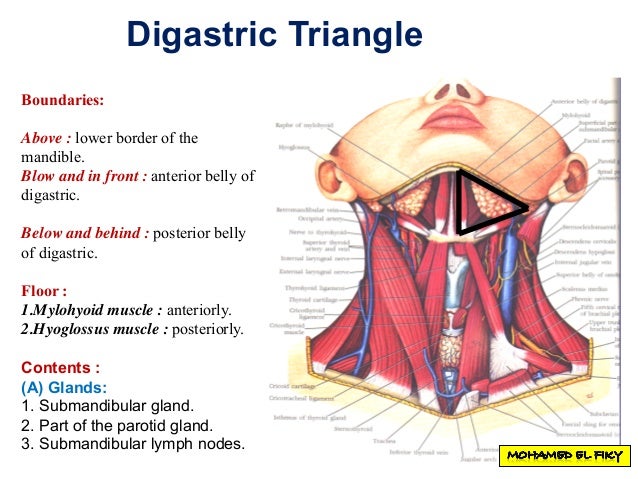
Floor Muscles Of Digastric Triangle
Digastric Or Submandibular Triangle Earth S Lab

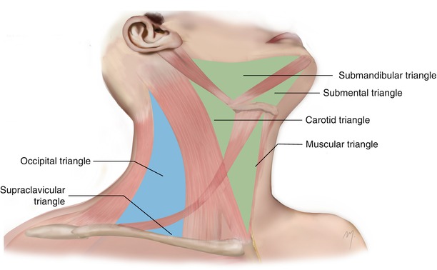
Anterior Triangle Of The Neck L4 Flashcards Quizlet

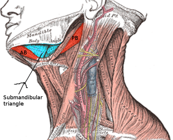
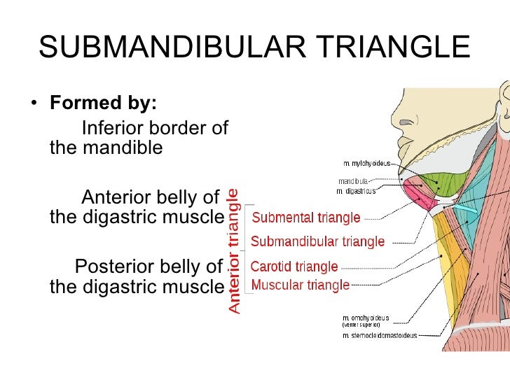
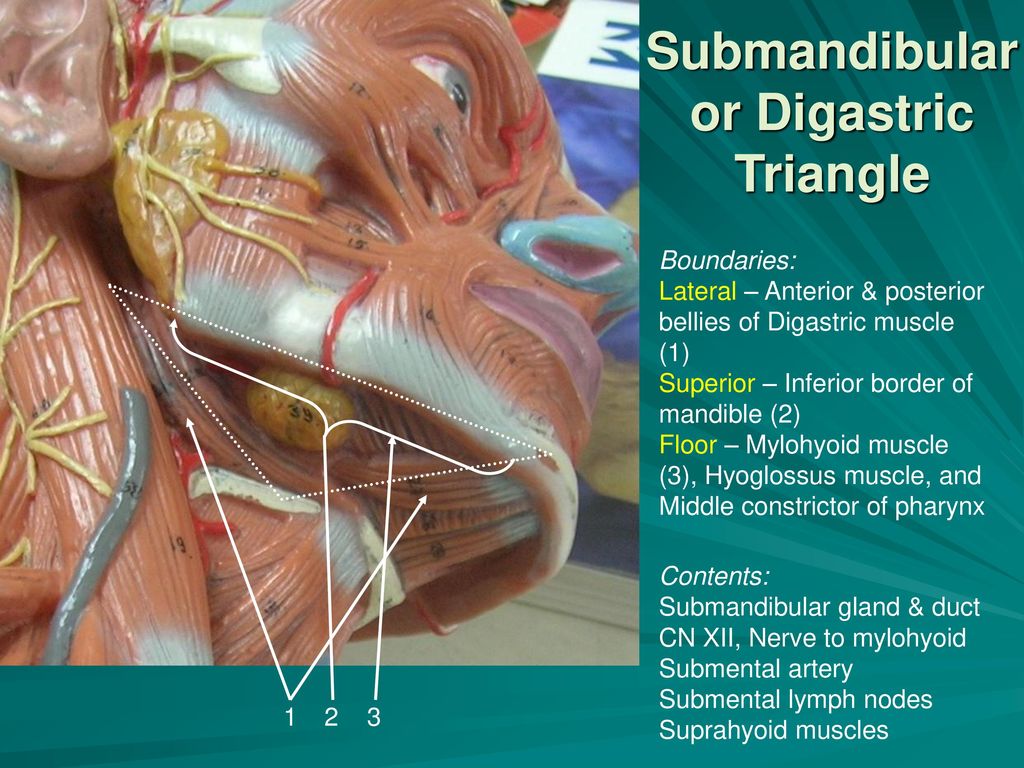
Contents Of Submandibular Triangle

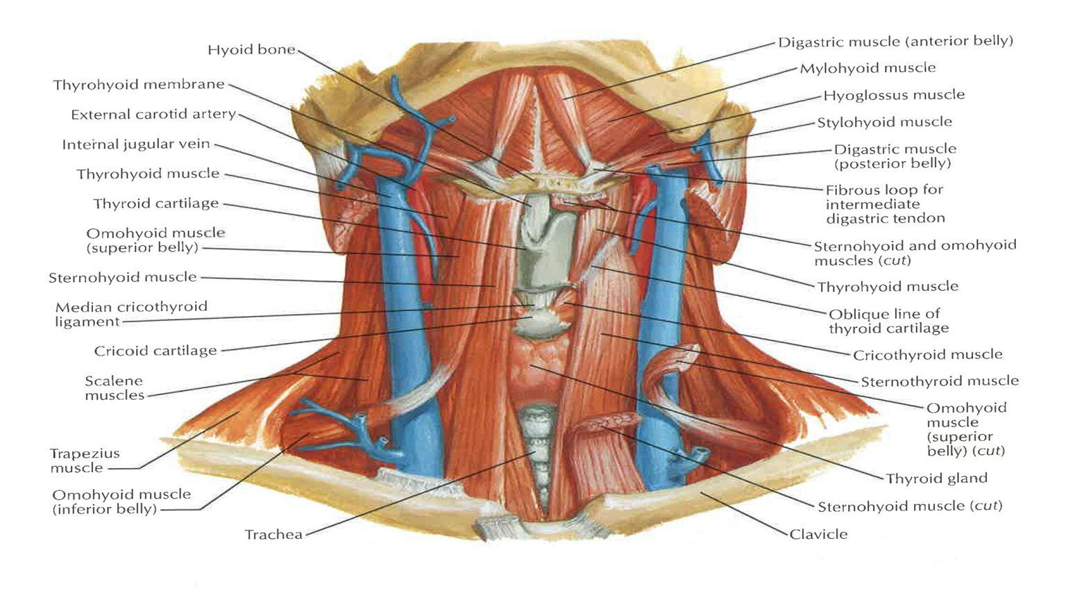
Anatomy Of The Neck By Dr Rasha Sabry Ppt Video Online Download

Digastric Triangle Boundaries And Contents Animated Gross Anatomy Head And Neck Youtube

The Submandibular Gland Structure Vasculature Innervation Teachmeanatomy
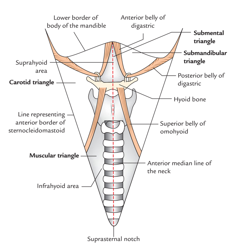
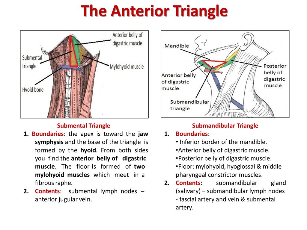
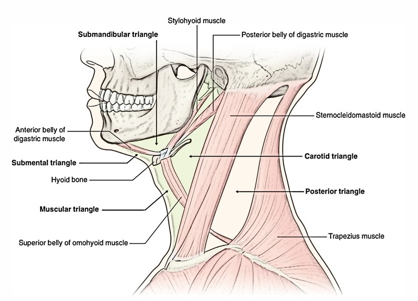
Digastric or submandibular triangle.
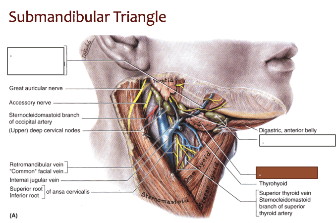
Floor muscles of digastric triangle.

Quiz6 Flashcards Quizlet

Jaypeedigital Ebook Reader
Anatomy Head And Neck Neck Triangle Article Statpearls
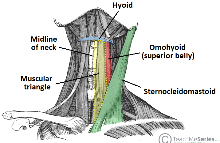
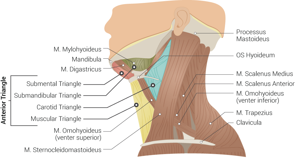
Anterior Triangle Of The Neck Subdivisions Teachmeanatomy
Anterior Triangle Of The Neck Part 1
Anatomy Head And Neck Submandibular Triangle Article Statpearls
Submandibular Triangle Anatomy And Clinical Notes Kenhub
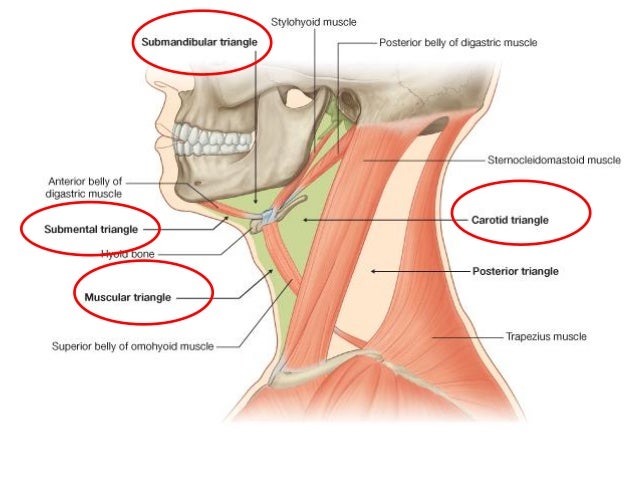
Triangles Of Neck By Dr Juveria Majeed Ms Ent
Easy Notes On Anterior Triangle Of The Neck Learn In Just 3 Minutes Earth S Lab
Triangles Of The Neck Ppt Year 1

Neck Triangles Flashcards Quizlet

Triangles Of The Neck Anatomy Borders And Contents Kenhub

Neck Anatomy E Lab
Unit Iv Problem Iv Anatomy Ppt Download
Anterior Triangle Of Neck
Anterior And Posterior Triangles Of Neck Ppt Download

Case Based Learning Triangles Of Neck Region
001b Superficial And Deep Structures Of The Neck Anatomy Ii Flashcards Memorang
Https Encrypted Tbn0 Gstatic Com Images Q Tbn 3aand9gct Pskepy Zarbqvusgf1ltdg526fna1yyiab Fuuhyxss Lkdy Usqp Cau
Easy 3 Mins Notes On Suprahyoid And Infrahyoid Muscles Of The Neck Earth S Lab
Https Www Medicinebau Com Uploads 7 9 0 4 79048958 Anatomy Ns Note 23 Pdf
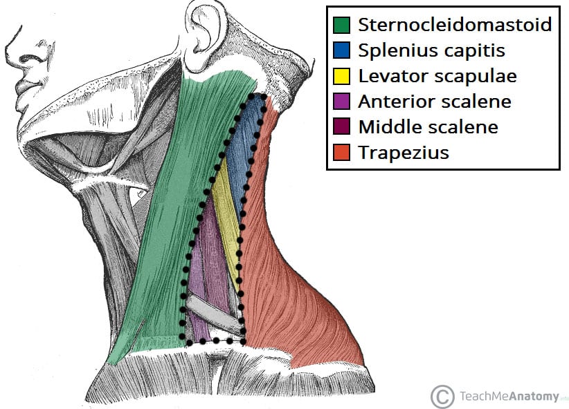
Posterior Triangle Of The Neck Subdivisions Teachmeanatomy
Neck 2 Anterior Triangle Head And Neck Anatomy Flashcards Memorang
Neck Plastic Surgery Key
Source : pinterest.com
