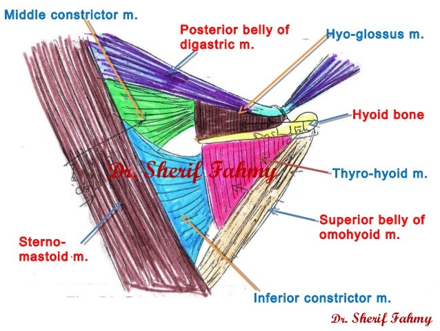
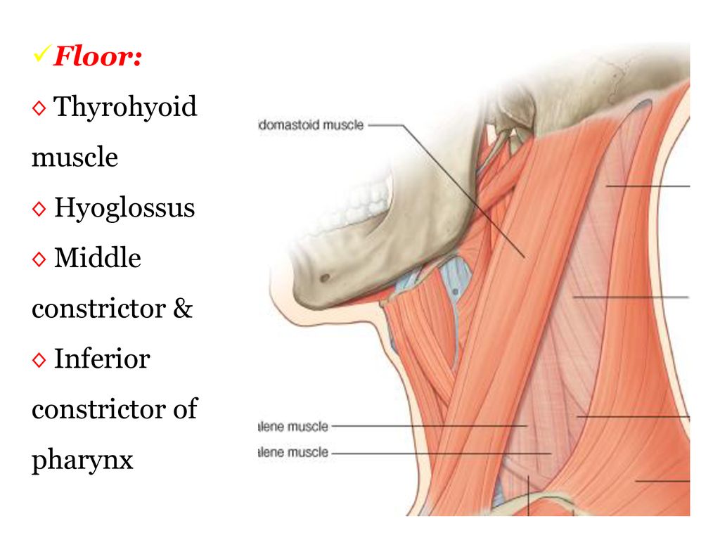
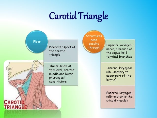
Floor Of Carotid Triangle

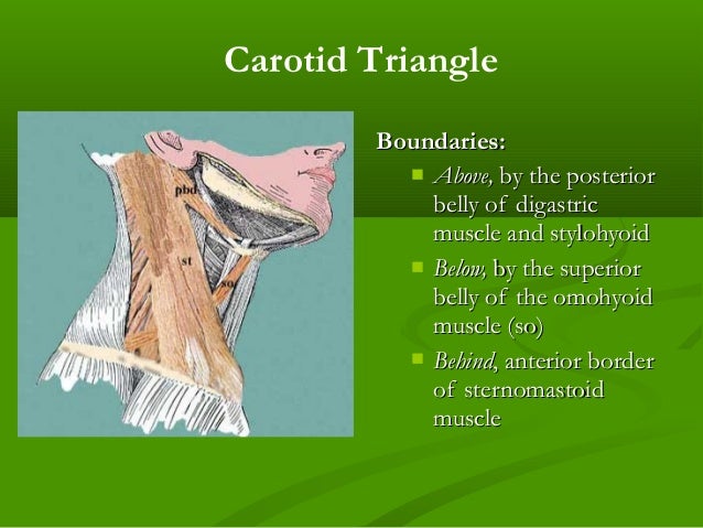
Carotid Triangle Boundaries Contents Anatomyqa
Carotid Triangle Anatomy Kenhub
Carotid Triangle Anatomy Of The Neck
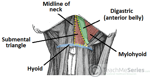
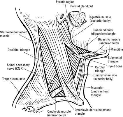
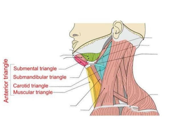
Anterior Triangle Of The Neck Subdivisions Teachmeanatomy

Jaypeedigital Ebook Reader

Jaypeedigital Ebook Reader
Structure superficial to mylohyoid in anterior digastric triangle is mylohyoid artery nerve.
Floor of carotid triangle.

Carotid Triangle Radiology Reference Article Radiopaedia Org

Carotid Triangle Boundaries Contents Anatomy Tutorial Youtube
Triangles Of Neck By Dr Juveria Majeed Ms Ent

Carotid Triangle Animated Gross Anatomy Head And Neck Medical Animation Youtube
Anterior Triangle Of The Neck Part 1

Case Based Learning Triangles Of Neck Region

Anatomy Pptx د سيف Muhadharaty
Anterior Triangle Of The Neck

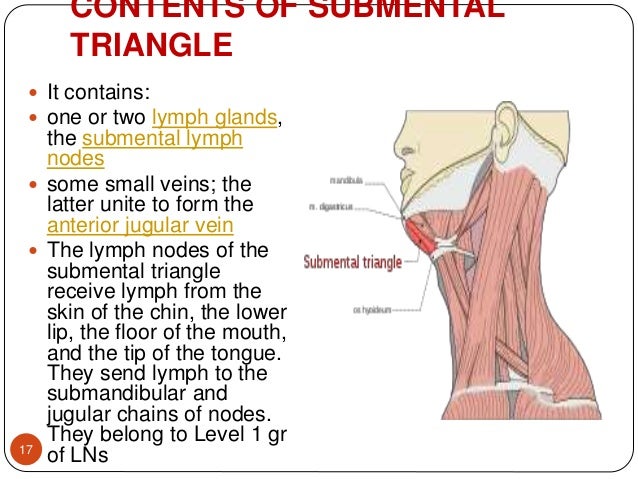
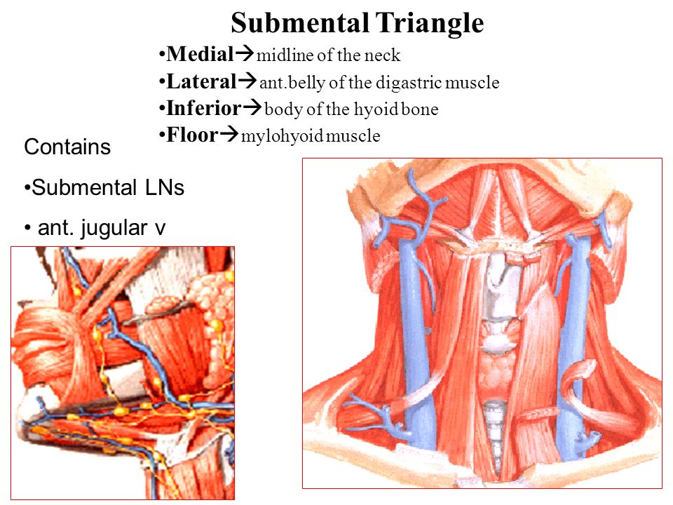
Contents Of Submandibular Triangle

Pdf Triangles Of The Neck A Review With Clinical Surgical Applications
Triangles Of The Neck

Triangles Of The Neck

Cb 724 Triangles Of The Head And Neck Flashcards Quizlet
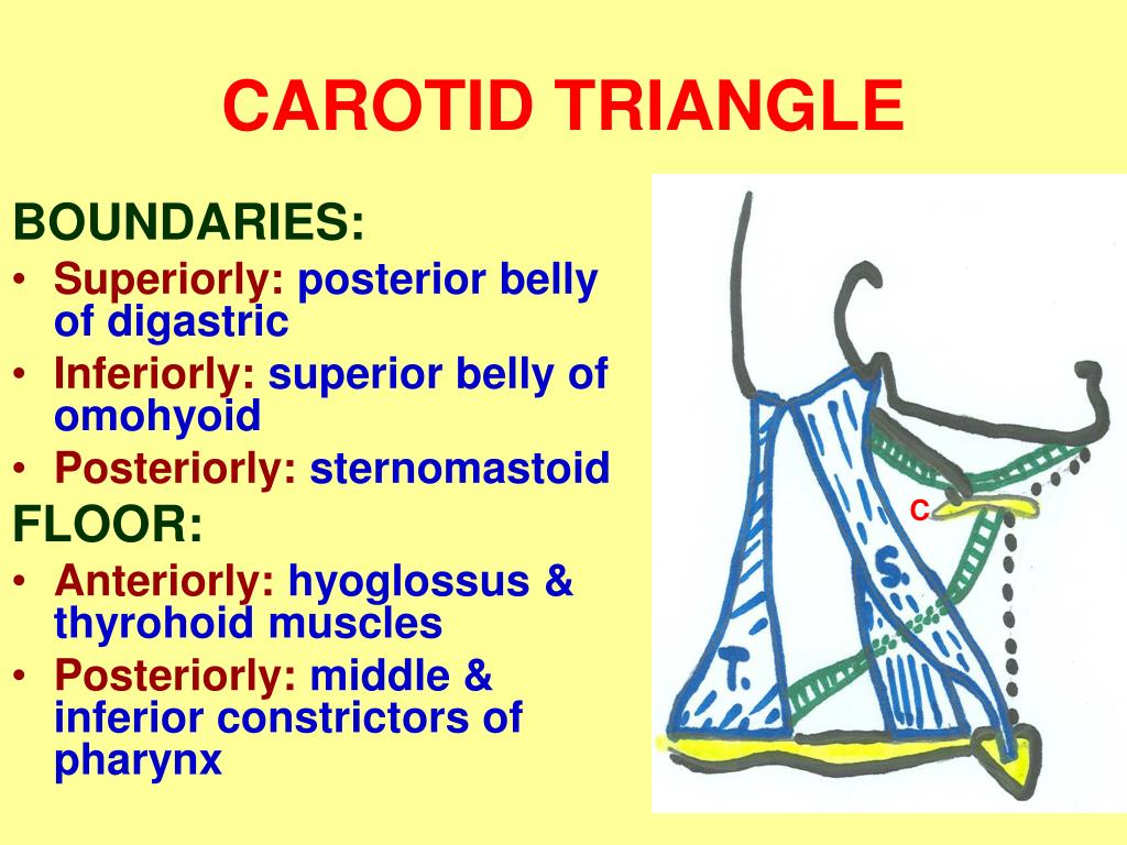
B Carotid Triangle Superiorly Posterior Belly Of Digastric Ppt Download
Suboccipital Triangle Ppt Video Online Download
Stylohyoid Origin Insertion Innervation And Action Kenhub
Surgical Anatomy Of Triangles Of Neck
Triangles Of The Neck
1
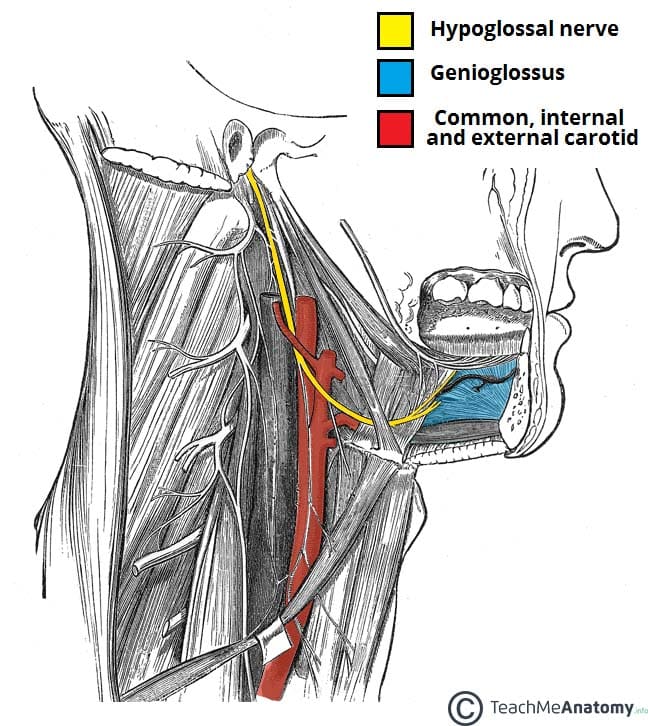
The Hypoglossal Nerve Cn Xii Course Motor Teachmeanatomy
Anterior Triangle Dr Lubna Nazli Associate Professor Anatomy Ppt Video Online Download

Triangles Of Neck Flashcards Quizlet

Subclavian Triangle Wikipedia
Ppt Anterior Triangle Of The Neck Ii Powerpoint Presentation Free Download Id 5351557
Source : pinterest.com
