Floor Of Lateral Cervical Region
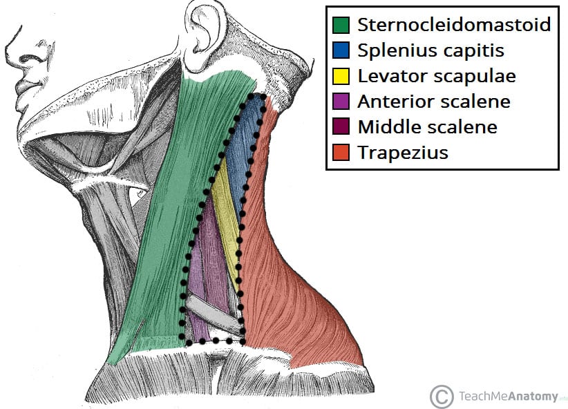
Posterior Triangle Of The Neck Subdivisions Teachmeanatomy
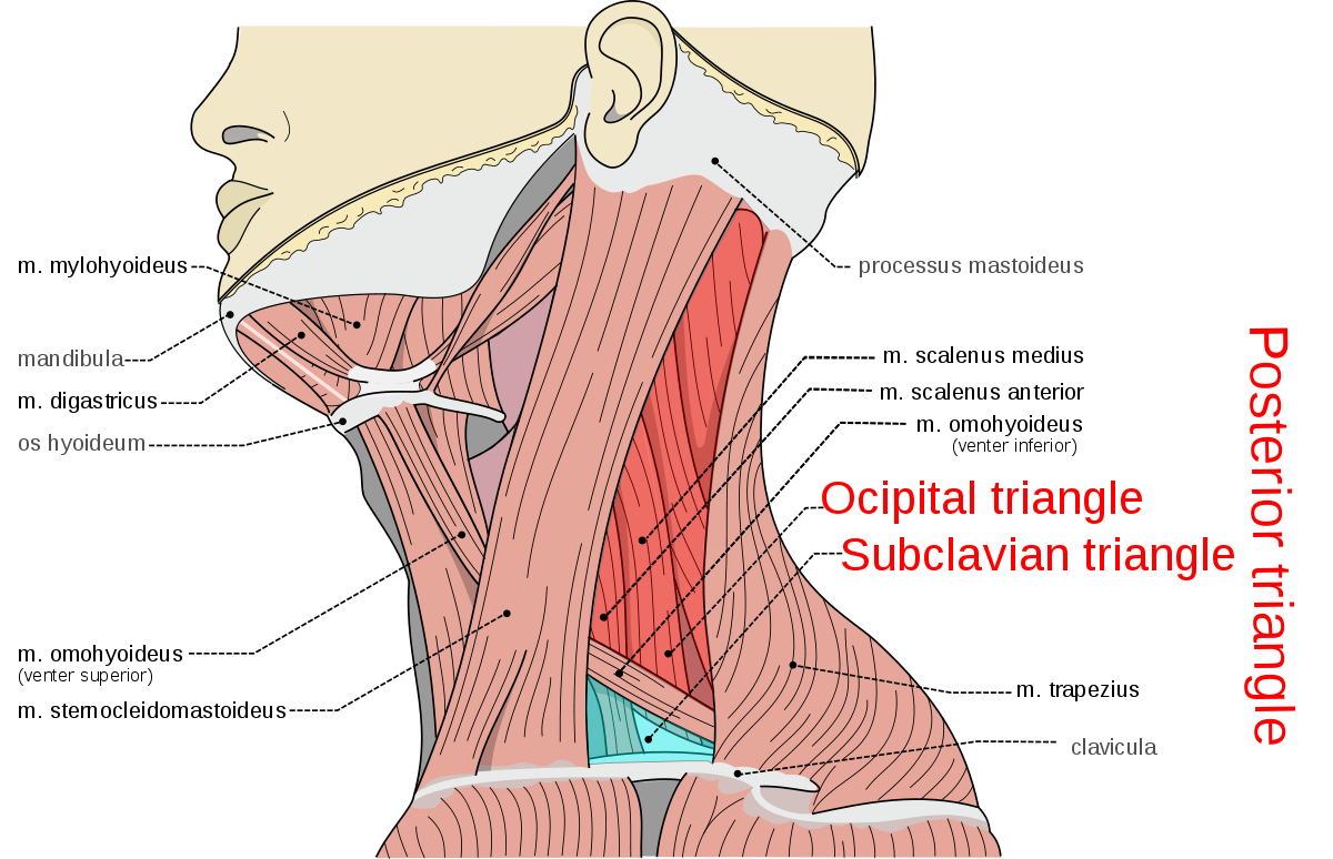
Posterior Triangle Of The Neck Wikipedia

Lateral Cervical Region Flashcards Quizlet

11 6 Axial Muscles Are Muscles Of The Head And Neck Vertebral Column Trunk And Pelvic Floor

Image Result For Anatomy Of Jaw And Ear Dental Anatomy Human Anatomy And Physiology Anatomy And Physiology

Gross Anatomy Of Neck Spencer Flashcards Quizlet

They are further divided into more specific groups based on a number of determinants.
Floor of lateral cervical region.

Opening Of The Mandible Anatomy Retrodiscal Tissue Bilaminar Zone Retrodiscal Tissue Bilaminar Zone Expanded Flo In 2020 Anatomy Medical Knowledge Body Anatomy

Floor Of Fourth Ventricle Note Both Vagal Hypoglossal Triangle Facial Nerve Vagus Nerve Brain Anatomy

Musculature Of Pharynx Much Of The Framework Of The Lateral And Posterior Walls Of The Pharynx Is Formed By An In 2020 Plexus Products Medical School Studying Muscle

Ligaments Of The Pelvic Region Beckenboden Anatomie Gesundheit Und Wellness
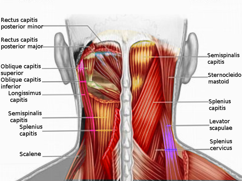
Anatomy Head And Neck Posterior Cervical Region Article Statpearls

Cervical Plexus1342596044233 Jpg 994 1010 Plexus Products Cervical Arteries Anatomy

Triangles Of The Neck Anatomy Borders And Contents Kenhub

Neck Atlas Of Anatomy

So How Do We Get It Back Here S A Good Place To Start Breathing Rolling Crawling Train Movements Not Muscles Lift Heavy Sh T W Anatomy Muscle Anatomy Muscle

Diaphragm Abdominal Surface

Muscle Attachments Of Skull Lateral View Norma Lateralis Human Skull Anatomy Anatomy Muscles Of The Face

Essential Anatomy 3 Anatomy App Anatomy Health And Fitness Apps

תוצאת תמונה עבור Cervical Spine Rom Goniometry Thoracic Region Cervical Vertebrae Thoracic

6 Simple Exercises To Build A Strong Core Pelvic Floor Transversus Abdominis Core Muscles

The Sternocleidomastoid Muscle And The Omohyoid Muscle Are Intersecting On The Side Of The Cervical Spine When The Sternocleidomastoid Muscle Muscle Cervical

Pin On Stretches For Hands And Neck

Orbit Walls Bones All Anatomy Bones Anatomy Human Brain Anatomy

Ap Neck Radiograph X Ray Radiology Student Radiology Medical Anatomy
1

The Scalenes The Dynamic Duo 1 Massage Therapy School Plexus Products Massage Therapy

Streptococcus Swollen Lymph Nodes Lymph Nodes Lymph Glands

Figure 9 9 Superficial Dissection Of Lateral Cervical Region

Nerves In Back Human Body Organs Anatomy Back Human Anatomy

Pin On Cervical Injection Exhibits
Source : pinterest.com
