Floor Of The Orbit Panoramic

Dentaltown Where The Dental Community Lives Dental Dental Hygenist Dentaltown

Anatomical Landmarks On Panoramic Radiography Dentstudy Com
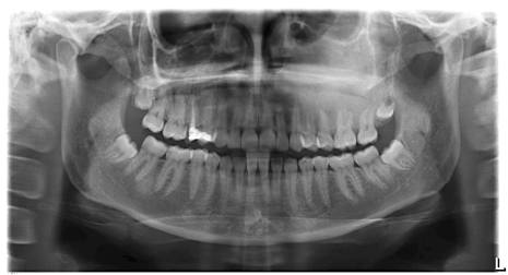
Figure 4

Limited Space Panoramic Outdoor Advertising Floor Graphics

Radiographic Anatomy Of The Skull Organizacao De Estudo

Panoramic Error Recognition Flashcards Quizlet

The forms the floor of the orbit of the eyes the sides and floor of the nasal cavity and the hard palate.
Floor of the orbit panoramic.

This Date In Science John Glenn First American To Orbit Earth Earthsky Org John Glenn Earth From Space Nasa Earth

Inner Orbit Lvov Left And Trocto Right By Hybycozo Interactive Art Mirror Modern

Everythingandsome Home House Design Penthouse Living
Https Onlinelibrary Wiley Com Doi Pdf 10 1111 J 1834 7819 2011 01655 X

Pin On Things To Do In London

This Modern New York Penthouse Features Panoramic Views And Sophisticated Decor Stylish Bathroom New York Apartment Sophisticated Decor

Squamous Cell Carcinoma Floor Of The Mouth Panoramic Radiograph Download Scientific Diagram

Panoramic Advertising Format For Warner Bros Pacific Rim At The Metro Centre Panoramic Metrocentre Mallad Outdoor Advertising Metro Center Floor Graphics

30 Modern Corner Windows For Framed And Frameless Panoramic Views Farmhouse Style House Plans Modern Farmhouse Floorplan Farmhouse Architecture

Jp London Md4a134 Solar System Space Removable Panoramic Https Www Amazon Com Dp B008592uh4 Ref Cm Sw R Pi Awdb X T Wall Murals Mural Panoramic

Arounder Travel And Lifestyle 360 Panoramas Castle Panorama Germany

After First Collaborating In 2011 And Again In 2012 Tetra Pak And Orbit Design Studio Reteamed A Third Time To Craft A New Tetra Pak Tetra Office Relocation

Artist S Concept Of Skylab Space Station Cluster In Earth S Orbit Original From Nasa Digitally Enhanced By Rawpixel F Space Station Spacecraft Earth Orbit

Opportunity Explored The Rim Of Endeavor Crater For Five Years The Rover Is Now Descending Toward The Crater S Floor Curiosity Mars Mars Surface Nasa

Pin On Office

Pin On Design 2

A New Four Level Home In San Francisco Includes A Panoramic Roof Top Deck Glass Shower Enclosures Bathroom Inspiration Bathroom Decor

Gallery Of Studiokca S Nasa Orbit Pavilion Lets Visitors Listen To The Sounds Of Space 9 Acoustic Architecture Parametric Architecture Pavilion Architecture
Https Encrypted Tbn0 Gstatic Com Images Q Tbn 3aand9gcscvh Kknyllecpwc5iug P Hxffa19qs2jon9osypblfgj4vu0 Usqp Cau

House Plans Modern Sims 51 Ideas Modern Style House Plans Sims House Plans House Plans

Earth Orbit Night City Lights From Space Asia Stock Footage Night City Earth Orbit City Lights At Night Night City Outdoor

China S Probe Sends Panoramic Image Of Moon S Far Side Science And Technology News The Far Side Latest Science News

Last Panoramic From Curiosity Rover On Mars Http Www Db Prods Net Marsroversimages Curiosity Html Sol17 Space And Astronomy Space Exploration

Mysterious X 37b Military Space Plane Caught On Camera Photo Secret Space Camera Photo Vandenberg Air Force Base
Source : pinterest.com
