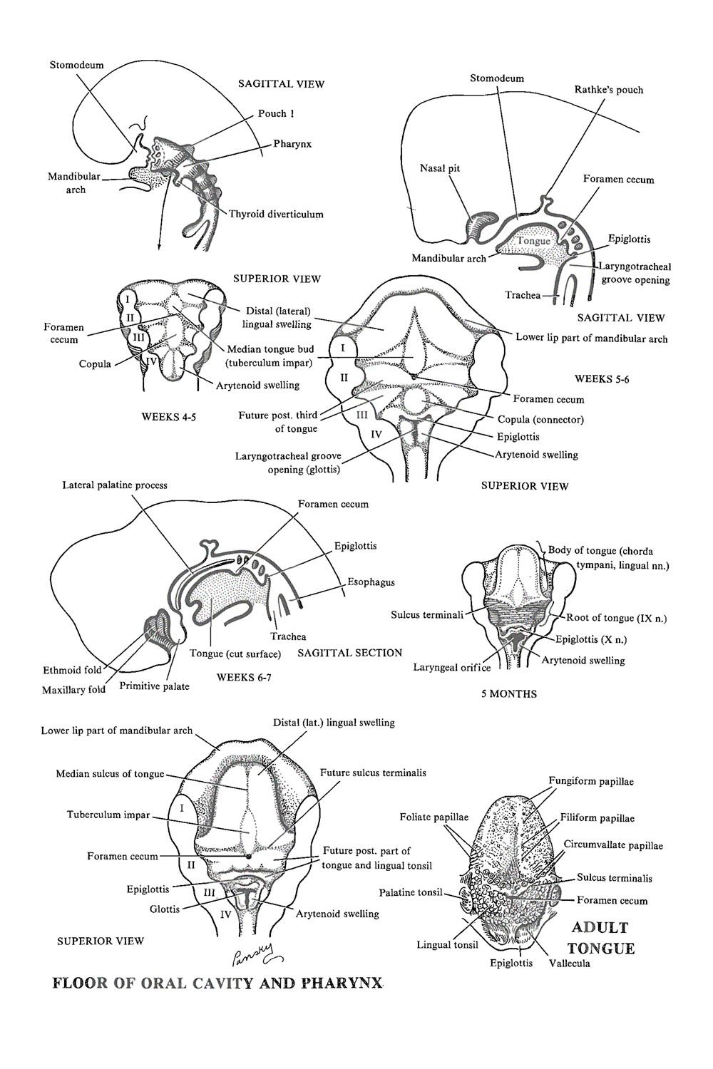
Floor Of The Pharynx
The Branchial Apparatus The Floor Of The Pharynx Tongue And Associated Structures Image 1 Mystic Messenger Mystic Messenger Comic Mystic Messenger Memes

Pharynx And Larynx Yahoo Image Search Results Thoracic Cavity Pearson Education Pulmonary Gas Exchange

Musculature Of Pharynx Much Of The Framework Of The Lateral And Posterior Walls Of The Pharynx Is Formed By An In 2020 Plexus Products Medical School Studying Muscle

The Muscles Of The Pharynx Medical Anatomy Human Anatomy And Physiology Anatomy And Physiology

Pharynx Subdivisions Muscles Nerve Supply And Applied Aspects Nerve Structure Maxillary Nerve Sensory Nerves

A Fun And Easy Way To Remember Laryngopharynx In Health Sciences Medical Anatomy Respiratory System Anatomy

Tongue largest single muscular organ in oral cavity.
Floor of the pharynx.
Instant Anatomy Head And Neck Areas Organs Mouth Tongue Development Head And Neck Body Anatomy Development

Flashcards Pharynx Medical Anatomy Shoulder Muscle Anatomy Health Science

Lymph Nodes Lymph Nodes Lymph Massage Upper Limb Anatomy

Pin On Medical

Lymph Nodes Of The Face And Neck What Causes Swollen Lymph Nodes Under The Collar Bone Lymphmassage Lymph Massage I 2020

Muscles Of Pharynx Lateral View Anatomy Pharyngobasilar Fascia Tensor Veli Palatini Muscle Levator Veli Palatini Anatomy Medical Anatomy Anatomy Images

Anatomy Anatomy And Physiology Anatomy Definition Anatomy Of The Foot Anatomy Of The Guts Ana Anatomy And Physiology Textbook Anatomy And Physiology Anatomy

Instant Anatomy Head And Neck Areas Organs Temporal Fossa Bones Head And Neck Body Anatomy Anatomy

Respiratory System Diagram Respiratory System Anatomy Respiratory System Anatomy Respiratory System Human Body Systems

Instant Anatomy Head And Neck Areas Organs Salivary Glands Submandibular Dental Hygiene School Medical Anatomy Human Anatomy And Physiology

Digestive System In 2020 Digestive System Human Digestive System Endocrine System

Respiratory System Respiratory System Human Respiratory System Respiratory

Instant Anatomy Head And Neck Muscles Palate Dental Anatomy Dental Assistant Study Dental Hygiene School

What Are Some Causes Of Dry Mouth Dry Mouth Dry Mouth Mouth Anatomy Dental Assistant

Parts Of Lips Mouth Oral Cavity Cancer Soft Palate Oral Cavity

Picturing Medicine Anatomy

Cranial Nerves Anatomy Clinical Signs And Study Tips Cranial Nerves Cranial Nerves Anatomy Nerve Anatomy

The Anatomy Of The Tongue Google Search
Https Encrypted Tbn0 Gstatic Com Images Q Tbn 3aand9gcrfbiurztv4scy Uc7na Wtvb8bznyxtswpl1utma7wxy5auqta Usqp Cau

Pin On G K Question

Pin On Medical

Craniofacial Development Development Soft Palate Ductus Arteriosus

Nuclei Functional Components And Distribution Of Cranial Nerves Textbook Of Clinical Neuroanatomy 2 Ed In 2020 Cranial Nerves Parotid Gland Cranial Nerves Mnemonic

Pin On Eten En Drinken
Source : pinterest.com
