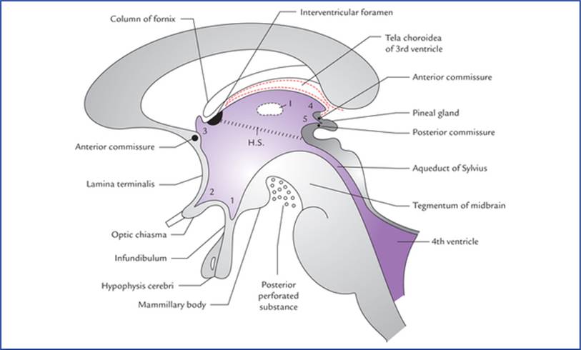
Floor Of Third Ventricle Perforation

Boundaries Of The Third Ventricle Plexus Products Medical Anatomy Boundaries
Third Ventricle Anatomy Kenhub

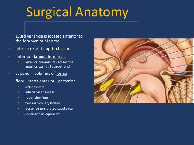
Approaches To Third Ventricular Tumors Sciencedirect
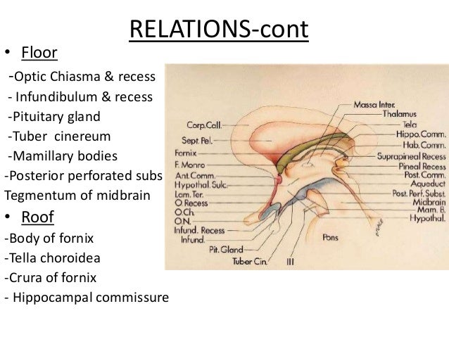
Diencephalon And Third Ventricle Textbook Of Clinical Neuroanatomy 2 Ed

Schematic Drawing Of The Floor Of The Third Ventricle Ideally The Download Scientific Diagram

Jaypeedigital Ebook Reader

Transient diabetes insipidus one of its rarest complications.
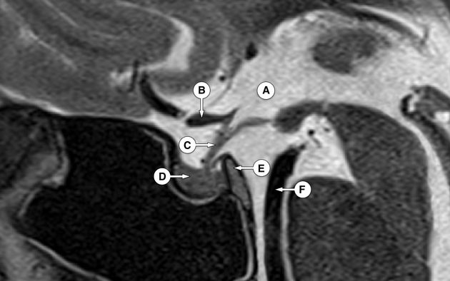
Floor of third ventricle perforation.

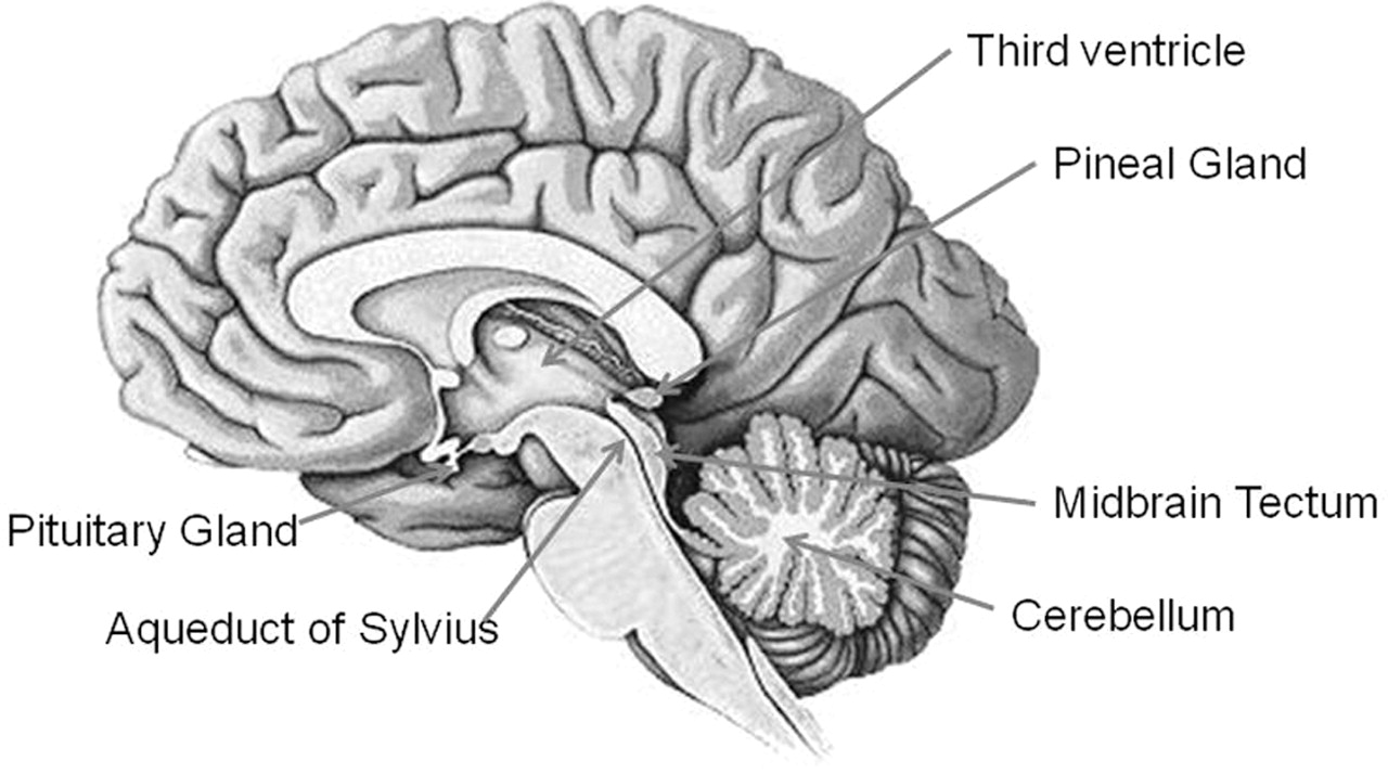
3rd Ventricle N Pineal Gland
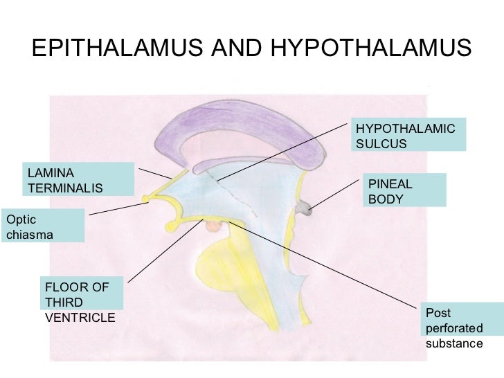
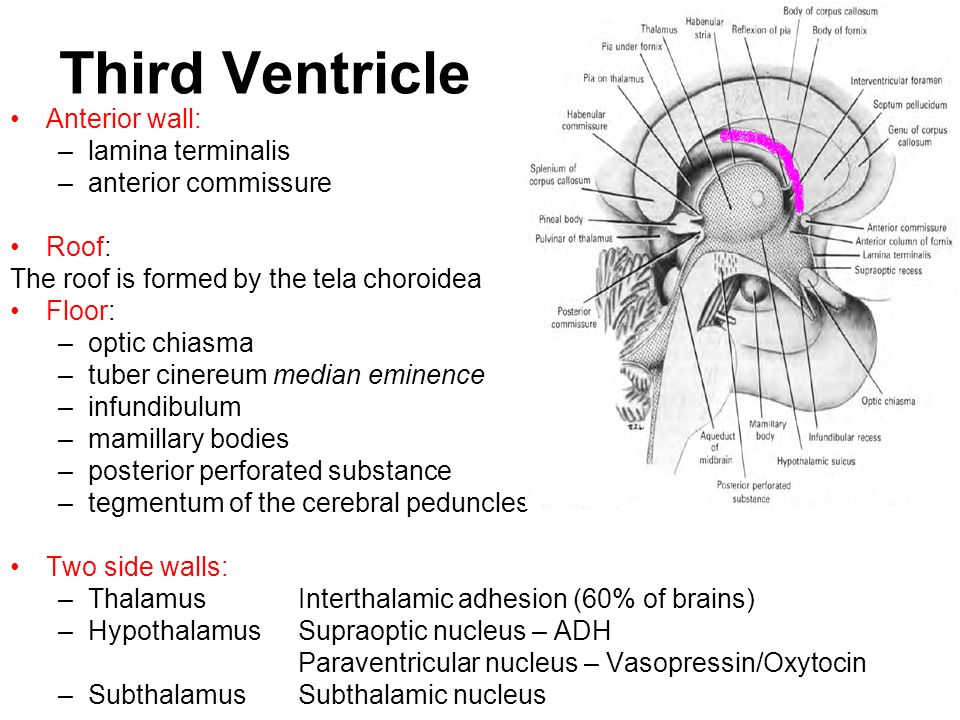
Third Ventricle

4 Lateral And Third Ventricle Anatomy Neupsy Key

The Third Ventricle Its Width And Height A Width Of The Third Download Scientific Diagram

Neurolab Exercise 3 Quiz Questions Post Lab Flashcards Quizlet
Third Ventricle
Plos One Exploring The Efficacy Of Endoscopic Ventriculostomy For Hydrocephalus Treatment Via A Multicompartmental Poroelastic Model Of Csf Transport A Computational Perspective
Floor Of Third Ventricle Mnemonics
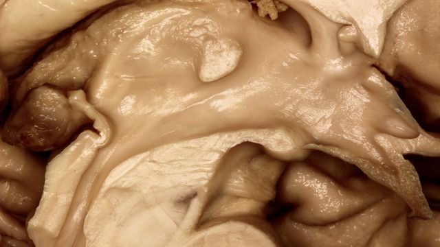
Floor And Roof Of The Third Ventricle Neuroanatomy The Neurosurgical Atlas By Aaron Cohen Gadol M D

Endoscopic Third Ventriculostomy Neupsy Key

Ventricles And Coverings Of The Brain Clinical Neuroanatomy 28 Ed
Brain Anatomy
Walter S Hydrocephalus Education Blog What Is Endoscopic Third Ventriculostomy Etv Part 1 Of 2
Http Bmcsagar Edu In New Upload Third 20ventricle Pdf

Pdf Histological Analysis Of The Third Ventricle Floor In Hydrocephalic And Nonhydrocephalic Brains Application To Neuroendocrine Complications Following Third Ventriculostomy Procedures Laboratory Investigation

Pdf Suprapineal Recess Diverticulum Of The Third Ventricle Exerting A Mass Effect On The Midbrain And Cerebellum
Floor Of 3rd Ventricle Form By Mnemonics

Jaypeedigital Ebook Reader
Https Encrypted Tbn0 Gstatic Com Images Q Tbn 3aand9gcqg Frvu7jo23x Tpbzylw36yxempobu31o3i7xumhvpr42vnaa Usqp Cau

Ajr Mobile Ulcers Peptic Ulcer Sonography
Meninges Csf Ventricular System Objectives Describe The Arrangement Of The Meninges And Their Relationship To Brain And Spinal Cord Explain The Occurrence Ppt Download

Ventricular Deformities Associated With Aqueductal Stenosis The Ispn Guide To Pediatric Neurosurgery

81 Third Ventricle الدكتور أحمد كمال المقطع الأخير Youtube
Third Ventricle Radiology Reference Article Radiopaedia Org
Source : pinterest.com
